
XCT Imaging
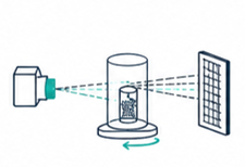
High-resolution X-ray computed tomography (XCT) for medical devices, biomaterials, hard tissues, and implants, enabling detailed internal structural analysis without sample destruction
Contrast-enhanced X-ray computed tomography (CECT)
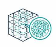
3D histology of soft biological tissues and organs, combining advanced contrast-enhancing staining agents (CESAs) with in-house synthesis, handling, and protocol optimisation to enable non-destructive, tissue-preserving 3D imaging
Cryogenic contrast-enhanced XCT (cryo-CECT)
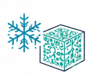
enabling high-resolution imaging under cryogenic conditions for improved contrast and enhanced preservation of fine microstructural features.
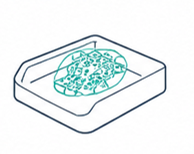
3D Imaging of Paraffin-Embedded Tissues
High-resolution 3D X-ray imaging of paraffin-embedded (FFPE) samples, enabling non-destructive volumetric analysis of tissues prepared using standard histopathology workflows.
Seamless integration with conventional histology, allowing correlation with 2D sections while preserving the full 3D structure of the sample

Data Processing and Analysis
Comprehensive 3D image processing and quantitative analysis of XCT, CECT, and cryo-CECT datasets for detailed investigation of tissue microstructure and biomaterial architecture
Multi-modal image correlation, integrating 3D imaging data with 2D microscopy or other modalities to provide a holistic understanding of biological structures

Reversible staining protocols, allowing removal of contrast agents and enabling subsequent analysis of the same sample using complementary techniques.
Classical 2D histology for complementary analysis
Consultancy services on CESA selection and preparation
Supply of proprietary non-destructive CESAs