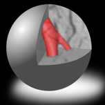
3D X-ray based histology is an emerging imaging technique to perform histology of biological tissues in full 3D and in a non-destructive manner. It is performed by imaging a sample with XCT and generating a 3D digital image dataset of the sample. During and after image acquisition, the sample remains intact and can be used for further evaluation, for instance with the classical 2D histology. The benefits of 3D X-ray-based histology extend beyond its non-destructive nature. Through high-resolution 3D imaging and data analysis, the technology also provides quantitative structural and compositional insights, making it highly applicable to longitudinal biomedical studies and (pre)clinical research. These advantages make it a powerful tool for advancing understanding of biological tissues and supporting various scientific and biomedical applications.


